PDF(51573 KB)
 PDF(51573 KB)
 PDF(51573 KB)
马尾松胚性愈伤组织对松材线虫的抗性评价
An in vitro evaluation of the resistance traits to pine wood nematode (Bursaphelenchus xylophilus) in Pinus massoniana embryogenic callus
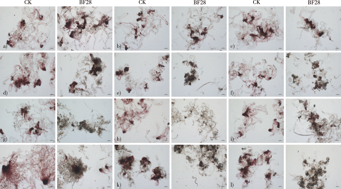
【目的】建立马尾松(Pinus massoniana)愈伤组织抗松材线虫(Bursaphelenchus xylophilus)病评价体系,筛选抗性马尾松细胞系。【方法】以马尾松不同胚性愈伤组织为试验材料,以黑松(P. thunbergii)和湿地松(P. elliottii)胚性愈伤组织作对照,接种无菌松材线虫,从接种线虫后愈伤组织外观形态、细胞结构和细胞活力变化以及线虫繁殖量4个方面探讨马尾松胚性愈伤组织对松材线虫的抗性。【结果】马尾松不同细胞系胚性愈伤组织之间抗病性差异显著。细胞系GX19-1-2和GX20-3-3显示出最高的抗性,接种无菌松材线虫10 d后外观轻微黄化,TTC染色结果为红色,显示其细胞保持较好活力;再分离线虫数量分别为(4 200±306)条和(5 933±1 392)条,显著低于湿地松2个细胞系(1907-9和1927-1),抗病性高于湿地松的1907-9和1927-1;GX20-3-8次之,线虫数量为(11 133±2 728)条;细胞系GX20-1-1接种无菌松材线虫后严重褐化、水渍化,TTC染色结果显示其细胞已完全失去活力,再分离线虫数量为(24 800±2 411)条,易感程度介于黑松细胞系1337和36-2之间。【结论】构建了基于愈伤组织的松树抗松材线虫评估体系,并筛选出2个具有较高抗性水平的马尾松细胞系GX19-1-2和GX20-3-3。这不仅为大规模筛选抗松材线虫病新种质奠定了基础,还为深入探究寄主松树-病原松材线虫之间相互作用的机制提供了新的研究平台。
【Objective】Pinus massoniana, a predominant tree species in Chinese forests, is highly susceptible to pine wood nematode (PWN), Bursaphelenchus xylophilus, which causes pine wilt disease and can lead to severe economic and ecological damage. This study aimed to develop an evaluation system for assessing the resistance of P. massoniana embryogenic callus to PWN and to screen for cell lines that exhibit enhanced resistance.【Method】The embryogenic callus induced from immature embryos of P. massoniana, sourced from Guangyun Forest Farm in Pingle County, Guilin City, Guangxi Zhuang Autonomous Region, China, was used as the experimental material, and the embryogenic callus of P. thunbergii and P. elliottii was used as the control group. The strongly virulent PWN strain AMA3c28, maintained at Nanjing Forestry University, China, was utilized for inoculation. Bacterial-free PWNs were obtained by sterilizing nematode eggs with 15% H2O2 for 50 min followed by rinsing in sterile water three times. The sterilized eggs were then inoculated onto pine callus, where they hatched and proliferated. The bacterial-free PWNs were collected and each callus piece from different cell lines was inoculated with 50 μL of a nematode suspension containing approximately 500 nematodes. Control groups were inoculated with an equal amount of sterile water. Post-inoculation, the cultures were incubated at 25 ℃ in the dark, and morphological changes were observed after 10 d, followed by the microscopic examination of cellular morphology. The cell viability was assessed using the 2,3,5-triphenyltetrazolium chloride (TTC) staining method. The population dynamics of PWN within the callus were evaluated by re-isolating the nematodes using the Baermann funnel technique. 【Result】The study revealed a marked variation in PWN resistance among the P. massoniana cell lines. The cell lines GX19-1-2 and GX20-3-3 exhibited the highest resistance and minimal morphological changes, maintaining an cell structural integrity after inoculation with bacterial-free PWNs for 10 days. In contrast, cell lines GX20-4-4, GX20-1-10, GX20-3-5, GX20-1-1 and GX20-1-7 exhibited severe browning, tortured cell structure, and significant growth inhibition, indicating weaker resistance. TTC staining confirmed these observations, with resistant cell lines showing vibrant red staining similar to the control groups, while susceptible lines turned pink and white, indicating reduced cell viability. Microscopic examination of cell structures post-inoculation further validated the resistance profiles, with resistant lines maintaining clear embryonic head-stem structures, and susceptible lines showing disrupted cellular integrity and a leakage of cellular contents. The population dynamics of PWN within the callus varied significantly among cell lines. Notably, the P. thunbergii cell line 36-2 exhibited the highest reproduction of PWN (107 333 ± 9 333), indicating a high susceptibility. In contrast, the P. massoniana cell lines GX19-1-2 and GX20-3-3 showed lower PWN levels, which were significantly lower than that of the two P. elliottii cell lines 1907-9 and 1927-1, suggesting a strong inhibitory effect on nematode reproduction and strong resistance. Cell line GX20-3-8 had similar resistance compared to that of P. elliotti cell lines. 【Conclusion】This study successfully developed an in vitro evaluation system for assessing the resistance of P. massoniana to PWN, revealing that cell lines GX19-1-2 and GX20-3-3 exhibit promising levels of resistance. The results act as a basis for future research, contribute to the development of resistant P. massoniana varieties, and can be employed to establish a new research platform for enhancing our understanding of the interactions between host pine trees and the PWN.
马尾松 / 松材线虫 / 胚性细胞系 / 细胞活力 / 线虫繁殖量 / 抗性评价
Pinus massoniana / Bursaphelenchus xylophilus / embryogenic cell lines / cell viability / nematode reproduction / resistance evaluation
| [1] |
理永霞, 张星耀. 松材线虫入侵扩张趋势分析[J]. 中国森林病虫, 2018, 37(5):1-4.
|
| [2] |
胡龙娇, 吴小芹. 松树抗松材线虫病机制研究进展[J]. 生命科学, 2018, 30(6):659-666.
|
| [3] |
张旭, 赵京京, 闫峻, 等. 2017年中国大陆松材线虫病灾害经济损失评估[J]. 北京林业大学学报, 2020, 42(10):96-106.
|
| [4] |
叶建仁. 松材线虫病在中国的流行现状、防治技术与对策分析[J]. 林业科学, 2019, 55(9):1-10.
|
| [5] |
李计顺, 潘佳亮, 刘超, 等. 2020年全国松材线虫病疫情流行情况分析[J]. 中国森林病虫, 2021, 40(4):1-4.
|
| [6] |
徐六一, 章健, 高景斌, 等. 安徽省松材线虫病抗性育种研究进展[J]. 安徽林业科技, 2013, 39(2):8-10,14.
|
| [7] |
|
| [8] |
郝焰平, 姜春武, 陈雪莲, 等. “皖抗6号” 等六个抗松材线虫病马尾松无性系选育[J]. 中国森林病虫, 2021, 40(3):14-20.
|
| [9] |
刘进平, 郑成木. 体外选择与体细胞无性系变异在抗病育种中的应用[J]. 遗传, 2002, 24(5):617-630.
|
| [10] |
|
| [11] |
|
| [12] |
陈婷婷, 叶建仁, 吴小芹, 等. 抗松材线虫病马尾松体胚发生与植株再生条件的优化[J]. 南京林业大学学报(自然科学版), 2019, 43(3):1-8.
|
| [13] |
季孔庶, 王潘潘, 王金铃, 等. 松科树种的离体培养研究进展[J]. 南京林业大学学报(自然科学版), 2015, 39(1):142-148.
|
| [14] |
吴静, 朱丽华, 许建秀, 等. 抗松材线虫病赤松胚性愈伤组织的诱导及增殖[J]. 南京林业大学学报(自然科学版), 2015, 39(1):17-21.
|
| [15] |
李淼, 檀根甲, 承河元, 等. 植物抗病性研究现状与前景展望[J]. 江西农业大学学报(自然科学版), 2002, 24(5):731-736.
|
| [16] |
张立钦, 李传道. 用组织培养方法鉴别植物的抗病性[J]. 浙江林学院学报, 1989, 6(3):88-95.
|
| [17] |
|
| [18] |
|
| [19] |
|
| [20] |
|
| [21] |
|
| [22] |
张立钦, 李传道, 黄敏仁. 杨树组织培养愈伤组织对水泡型溃疡病的抗性[J]. 南京林业大学学报, 1989, 13(4):9.
|
| [23] |
|
| [24] |
林雪坚, 吴光金, 程淑华, 等. 桉树愈伤组织抗病性的测定及其快速繁殖研究[J]. 中南林学院学报, 2003, 23(6):117-120.
|
| [25] |
高立宏, 汉丽萍, 辛树权. 稻瘟病菌粗毒素对水稻成熟胚愈伤组织诱导的影响及抗性筛选[J]. 长春师范学院学报(自然科学版), 2007, 26(5):72-76.
|
| [26] |
|
| [27] |
|
| [28] |
|
| [29] |
|
| [30] |
|
| [31] |
林丽, 周蕾, 潘珺, 等. 无菌和带菌松材线虫对赤松的致病性[J]. 林业科学, 2017, 53(5):82-87.
|
| [32] |
朱丽华, 季锦衣, 吴小芹, 等. 一种制备无菌松材线虫的方法[J]. 东北林业大学学报, 2011, 39(6):65-67,71.
|
| [33] |
刘华, 梅兴国. TTC法测定红豆杉细胞活力[J]. 植物生理学通讯, 2001, 37(6):537-539.
|
| [34] |
理永霞, 王璇, 刘振凯, 等. 松材线虫致病机理研究进展[J]. 中国森林病虫, 2022, 41(3):11-20.
|
| [35] |
叶建仁, 吴小芹. 松材线虫病研究进展[J]. 中国森林病虫, 2022, 41(3):1-10.
|
| [36] |
|
| [37] |
|
| [38] |
|
| [39] |
|
| [40] |
|
| [41] |
|
| [42] |
朱丽华, 章欣月, 夏馨蕊, 等. 无细菌松材线虫对马尾松的致病性[J]. 林业科学, 2020, 56(7):63-69.
|
| [43] |
王华光, 李良, 巨云为, 等. 鞭毛蛋白毒素导致的黑松超微结构病理学变化[J]. 南京林业大学学报(自然科学版), 2018, 42(6):137-144.
|
| [44] |
|
| [45] |
|
| [46] |
|
| [47] |
张艺, 吴小芹, 王钰, 等. 抗松材线虫病赤松家系的抗性测定及组织病理学观察[J]. 南京林业大学学报(自然科学版), 2007, 31(4):110-114.
|
| [48] |
李清清, 叶建仁, 朱丽华, 等. 抗松材线虫病赤松组培苗的抗病性测定1)[J]. 东北林业大学学报, 2013, 41(7):45-47.
|
| [49] |
吴小芹, 张艺, 陈蔚诗, 等. 黑松13个抗病家系对松材线虫的抗性反应及组织病理学观察[J]. 植物病理学报, 2008, 38(1):44-50.
|
| [50] |
|
| [51] |
徐福元, 葛明宏, 张培, 等. 不同马尾松种源对松材线虫病的抗病性[J]. 南京林业大学学报(自然科学版), 2000, 24(4):85-88.
|
/
| 〈 |
|
〉 |