 PDF(6586 KB)

The basic characteristics of stamen morphogenesis and development in Michelia figo
LIU Sitong, NIE Tangjie, WU Qingxian, JIN Leni, WAN Xiaoxia, YIN Zengfang
Journal of Nanjing Forestry University (Natural Sciences Edition) ›› 2024, Vol. 48 ›› Issue (6) : 34-40.
PDF(6586 KB)
 PDF(6586 KB)
The basic characteristics of stamen morphogenesis and development in Michelia figo
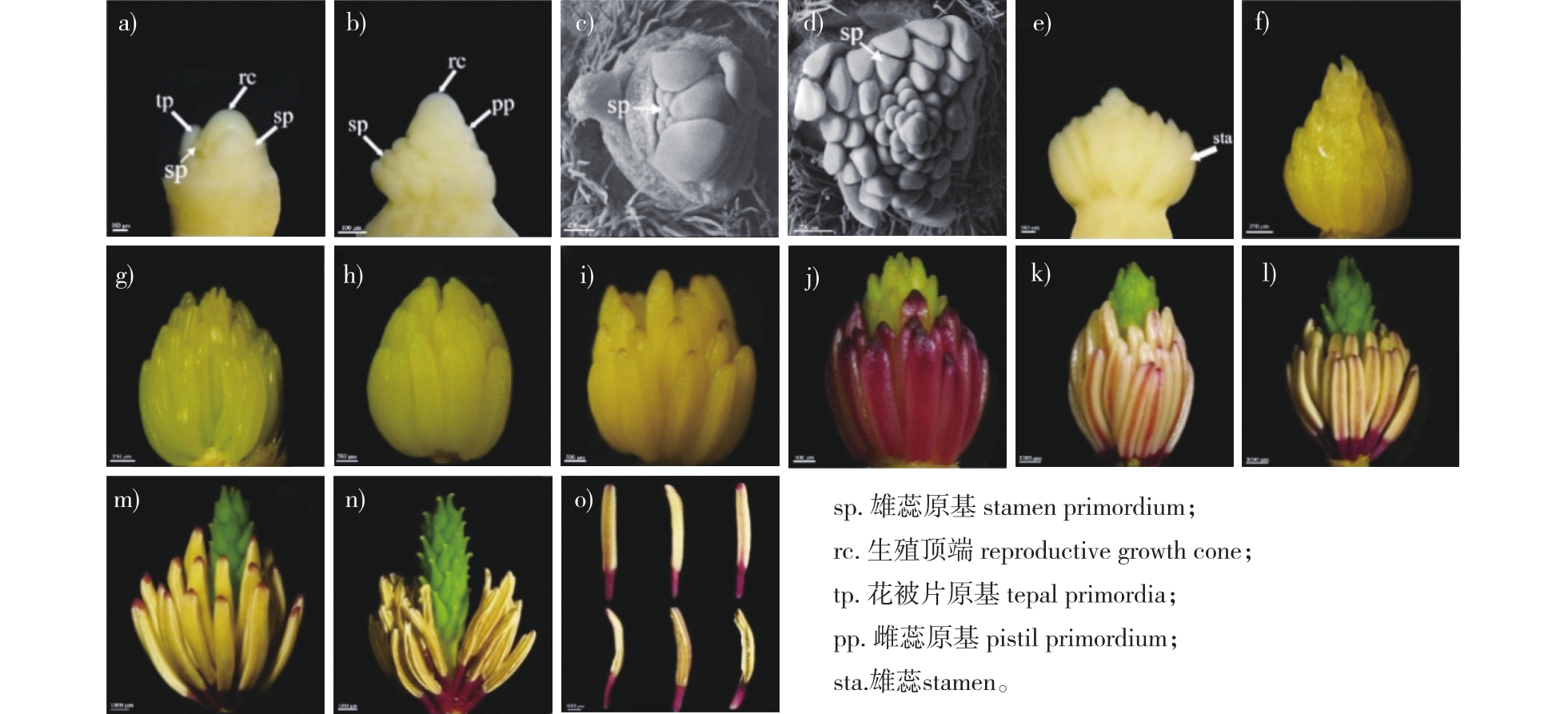
【Objective】The fundamental characteristics of stamen morphogenesis and development were described according to the observation of stamen morphogenesis in Michelia figo. The findings provide valuable basic data for future investigations in plant breeding and evolutionary biology within the Magnoliaceae family.【Method】Using the scanning electron microscopy and paraffin section technology, the stamens of flower buds in M. figo at different growth stages were observed.【Result】The formation pattern of stamen primordium in M. figo was found to be helical and centripetal. Upon the completion of stamen morphogenesis, distinct morphological changes were observed, leading to the formation of a longer anther and a shorter filament. The anther tissue consists of anther septa and four anther locules. Normally, the sporogenous tissue differentiates gradually into microspore mother cells in the anther locule. The microspore mother cell meiosis was not synchronized, and eventually the isobilateral and tertrahedroid microspore tetrads were formed. Then, the free microspore developed into two-celled pollen and a few three-celled pollen. The structure of the anther wall was composed of one layer of epidermis cells, one layer of endothecium cells, two to three layers of middle layer cells, and one layer of secretory tapetum cells. During the development of mature pollen, the tapetum cells disintegrated and disappeared, but some middle layer cells persisted until the anther wall underwent dehiscence. The filaments were surrounded by a single layer of epidermis cells on the outer surface. Internally, the filaments were composed of parenchyma tissue, and a vascular bundle ran through the center toward the connective tissue. There was the cavity structure in the parenchyma of the filaments during the earlier development stage, which later disappeared during the mature stage of the stamen.【Conclusion】The process of stamen morphorgenesis in M. figo is normal compared with other Michelia plant species. The fundamental characteristics observed, including the helical centripetal formation of stamens, asynchronous meiotic division of microspore mother cells, temporal changes of the cavity structure in filaments, predominance of isobilateral tetrads, and two-celled pollen reflected the inherent nature of the Magnoliaceae family.
Michelia figo / stamen morphorgenesis / filament / anther / microspore tetrad
| [1] |
|
| [2] |
|
| [3] |
|
| [4] |
|
| [5] |
|
| [6] |
张风娟, 徐兴友, 陈凤敏, 等. 天女木兰小孢子发生及雄配子体发育的观察[J]. 经济林研究, 2008, 26(4):71-75.
|
| [7] |
陈丽园, 桑子阳, 陈发菊, 等. 红花玉兰大小孢子发生及雌雄配子体发育的研究[J]. 西北农林科技大学学报(自然科学版), 2016, 44(9):181-185.
|
| [8] |
王姗, 沈永宝, 鲍华鹏, 等. 宝华玉兰大小孢子发生和雌雄配子体发育过程中解剖结构的变化[J]. 植物资源与环境学报, 2021, 30(3):46-53.
|
| [9] |
潘丽琴, 郝建, 徐建民, 等. 灰木莲花药结构和花粉发育特征[J]. 林业科学研究, 2021, 34(6):107-113.
|
| [10] |
熊海燕, 刘志雄. 深山含笑大、小孢子发生和雌、雄配子体发育研究[J]. 植物研究, 2018, 38(2):212-217.
|
| [11] |
|
| [12] |
|
| [13] |
|
| [14] |
张冬莲, 念波, 汪志威, 等. 不同加工工艺对含笑花茶品质的影响[J]. 中国茶叶加工, 2019(1):31-36.
|
| [15] |
|
| [16] |
|
| [17] |
亓白岩, 周冬琴, 於朝广, 等. 8种含笑属植物的抗寒性研究[J]. 江苏农业科学, 2010, 38(5):258-263.
|
| [18] |
樊光毅, 胡烈栋. 含笑花苗培育技术探讨[J]. 园艺与种苗, 2018, 38(12):18-19.
|
| [19] |
|
| [20] |
周瑾, 柏永清, 曾妍, 等. 含笑花花粉萌发和花粉管生长的离体培养研究[J]. 湖北农业科学, 2022, 61(19):72-77.
|
| [21] |
|
| [22] |
|
| [23] |
|
| [24] |
付琳, 曾庆文, 徐凤霞, 等. 观光木的花器官发生[J]. 热带亚热带植物学报, 2007, 15(1):30-34.
|
| [25] |
|
| [26] |
万小霞. 三种木兰科植物分枝成花过程及其温度适应性[D]. 南京: 南京林业大学, 2021.
|
| [27] |
|
| [28] |
|
| [29] |
|
| [30] |
王利琳, 胡江琴, 庞基良, 等. 凹叶厚朴大、小孢子发生和雌、雄配子体发育的研究[J]. 实验生物学报, 2005(6): 490-500.
|
| [31] |
赵兴峰, 孙卫邦, 杨华斌, 等. 极度濒危植物西畴含笑的大小孢子发生及雌雄配子体发育[J]. 云南植物研究, 2008, 30(5):549-556.
|
| [32] |
付琳, 徐凤霞, 曾庆文, 等. 广西含笑的小孢子发生及雄配子体形成的研究[J]. 广西植物, 2011, 31(3):312-317,311.
|
| [33] |
敖成齐. 含笑小孢子的发生、雄配子体的发育及其系统学意义[J]. 广西植物, 2007, 27(6):836-839.
|
| [34] |
|
| [35] |
|
| [36] |
|
| [37] |
刘晨妮. 玉兰与含笑杂交生物学基础研究[D]. 南京: 南京林业大学, 2018.
|
/
| 〈 |
|
〉 |


